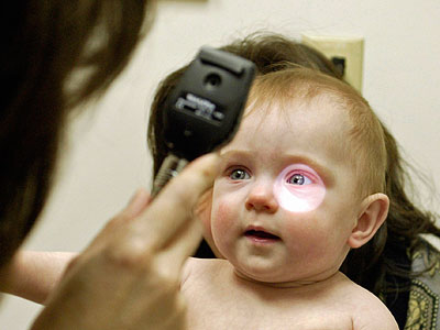
Computer-aided diagnosis of rare genetic disorders from family snaps
Computer analysis of photographs could help doctors diagnose which condition a child with a rare genetic disorder has, say Oxford University researchers.
The researchers, funded in part by the Medical Research Council (MRC), have come up with a computer programme that recognises facial features in photographs; looks for similarities with facial structures for various conditions, such as Down’s syndrome, Angelman syndrome, or Progeria; and returns possible matches ranked by likelihood.
Using the latest in computer vision and machine learning, the algorithm increasingly learns what facial features to pay attention to and what to ignore from a growing bank of photographs of people diagnosed with different syndromes.
The researchers report their findings in the journal eLife. The study was funded by the MRC, the Wellcome Trust, the National Institute for Health Research (NIHR) Oxford Biomedical Research Centre (BRC) and the European Research Council (ERC VisRec).
While genetic disorders are each individually rare, collectively these conditions are thought to affect one person in 17. Of these, a third may have symptoms that greatly reduce quality of life. However, most people fail to receive a genetic diagnosis.
‘A diagnosis of a rare genetic disorder can be a very important step. It can provide parents with some certainty and help with genetic counselling on risks for other children or how likely a condition is to be passed on,’ says lead researcher Dr Christoffer Nellåker of the MRC Functional Genomics Unit at the University of Oxford. ‘A diagnosis can also improve estimates of how the disease might progress, or show which symptoms are caused by the genetic disorder and which are caused by other clinical issues that can be treated.’
 The team of researchers at the University of Oxford included first author Quentin Ferry, a DPhil research student, and Professor Andrew Zisserman of the Department of Engineering Science, who brought expertise in computer vision and machine learning.
Professor Zisserman says: ‘It is great to see such an inventive and beneficial use of modern face representation methods.’
Identifying a suspected developmental disorder tends to require clinical geneticists to come to a conclusion based on facial features, follow up tests and their own expertise. It’s thought that 30–40% of rare genetic disorders involve some form of change in the face and skull, possibly because so many genes are involved in development of the face and cranium as a baby grows in the womb.
The researchers set out to teach a computer to carry out some of the same assessments objectively.
They developed a programme that – like Google, Picasa and other photo software – recognises faces in ordinary, everyday photographs. The programme accounts for variations in lighting, image quality, background, pose, facial expression and identity. It builds a description of the face structure by identifying corners of eyes, nose, mouth and other features, and compares this against what it has learnt from other photographs fed into the system.
The algorithm the researchers have developed sees patients sharing the same condition automatically cluster together.
The computer algorithm does better at suggesting a diagnosis for a photo where it has previously seen lots of other photos of people with that syndrome, as it learns more with more data.
Patients also cluster where no documented diagnosis exists, potentially helping in identifying ultra-rare genetic disorders.
‘A doctor should in future, anywhere in the world, be able to take a smartphone picture of a patient and run the computer analysis to quickly find out which genetic disorder the person might have,’ says Dr Nellåker.
‘This objective approach could help narrow the possible diagnoses, make comparisons easier and allow doctors to come to a conclusion with more certainty.’
###
Notes to Editors
The paper ‘Diagnostically-relevant facial gestalt information from ordinary photos’ by Quentin Ferry and colleagues is to be published in the journal eLife on Tuesday 24 January 2014.
The study was funded by the Medical Research Council, the Wellcome Trust, the National Institute for Health Research (NIHR) Oxford Biomedical Research Centre (BRC) and the European Research Council (ERC VisRec).
The Medical Research Council has been at the forefront of scientific discovery to improve human health. Founded in 1913 to tackle tuberculosis, the MRC now invests taxpayers’ money in some of the best medical research in the world across every area of health. Twenty-nine MRC-funded researchers have won Nobel prizes in a wide range of disciplines, and MRC scientists have been behind such diverse discoveries as vitamins, the structure of DNA and the link between smoking and cancer, as well as achievements such as pioneering the use of randomised controlled trials, the invention of MRI scanning, and the development of a group of antibodies used in the making of some of the most successful drugs ever developed. Today, MRC-funded scientists tackle some of the greatest health problems facing humanity in the 21st century, from the rising tide of chronic diseases associated with ageing to the threats posed by rapidly mutating micro-organisms. http://www.mrc.ac.uk
The National Institute for Health Research (NIHR) is funded by the Department of Health to improve the health and wealth of the nation through research. Since its establishment in April 2006, the NIHR has transformed research in the NHS. It has increased the volume of applied health research for the benefit of patients and the public, driven faster translation of basic science discoveries into tangible benefits for patients and the economy, and developed and supported the people who conduct and contribute to applied health research. The NIHR plays a key role in the Government’s strategy for economic growth, attracting investment by the life-sciences industries through its world-class infrastructure for health research. Together, the NIHR people, programmes, centres of excellence and systems represent the most integrated health research system in the world. For further information, visit the NIHR website.
Oxford University’s Medical Sciences Division is one of the largest biomedical research centres in Europe, with over 2,500 people involved in research and more than 2,800 students. The University is rated the best in the world for medicine, and it is home to the UK’s top-ranked medical school.
From the genetic and molecular basis of disease to the latest advances in neuroscience, Oxford is at the forefront of medical research. It has one of the largest clinical trial portfolios in the UK and great expertise in taking discoveries from the lab into the clinic. Partnerships with the local NHS Trusts enable patients to benefit from close links between medical research and healthcare delivery.
A great strength of Oxford medicine is its long-standing network of clinical research units in Asia and Africa, enabling world-leading research on the most pressing global health challenges such as malaria, TB, HIV/AIDS and flu. Oxford is also renowned for its large-scale studies which examine the role of factors such as smoking, alcohol and diet on cancer, heart disease and other conditions.
###
Press Office
.(JavaScript must be enabled to view this email address)
44-186-528-0530
University of Oxford