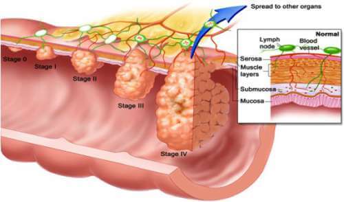
Being on the lookout for certain features of polyps may help physicians keep a closer eye on patients at risk for colorectal cancer.
Starting at age 50, or earlier with certain risk factors, patients are advised to be screened for colon cancer at regular intervals. Colonoscopy is an effective screening test because it allows doctors to find and view individual polyps (growths), and to remove them before they become cancerous.
Adenomas are polyps (small growths in the lining of the colon) that can vary in their size and shape, but are potentially precursors to colon cancer. Removal of these polyps reduces the risk of colon cancer. Flat adenomas are precancerous polyps that do not have a typical polyp- like appearance during endoscopy.
A new study in GIE: Gastrointestinal Endoscopy, the journal of the American Society for Gastrointestinal Endoscopy (ASGE), “Prevalence of advanced histological features and synchronous neoplasia in patients with flat adenomas,” indicates that a patient who had at least one flat adenoma had a higher chance of having multiple lesions with more advanced changes.
 The researchers looked at data from three clinical trials conducted at two medical centers that included patients undergoing screening or surveillance colonoscopy. The location, size, form and structure of each removed polyp was documented and sent for microscopic examination.
Risk Factors for Colorectal Cancer
The exact cause of colorectal cancer is not known, there are some factors that increase a person’s risk of developing the disease. These include:
- Age. The risk of developing colorectal cancer increases with age. The disease is most common in people over age 50, and the chance of getting colorectal cancer increases with each decade. However, colorectal cancer can develop in younger people.
- Gender. The risk overall is equal, but women have a higher risk for colon cancer, while men are more likely to develop rectal cancer.
- Polyps. Polyps are non-cancerous growths on the inner wall of the colon or rectum. While they are fairly common in people over age 50, one type of polyp, referred to as an adenoma, increases the risk of developing colorectal cancer. Adenomas are non-cancerous polyps that are considered precursors, or the first step toward colon and rectal cancer.
- Personal history. Research shows that women who have a history of ovarian, uterine, or breast cancer have a somewhat higher risk of developing colorectal cancer. A person who already has had colorectal cancer may develop the disease a second time, especially if the first disease was diagnosed before age 60. In addition, people who have chronic inflammatory conditions of the colon, such as ulcerative colitis or Crohn’s disease, are at higher risk of developing colorectal cancer.
- Family history. Parents, siblings, and children of a person who has had colorectal cancer are more likely to develop colorectal cancer themselves. If two or more family members have had colorectal cancer, the risk increases to about 20%. A family history of familial adenomatous polyposis, MYH-associated polyposis, or hereditary non-polyposis colon cancer (HNPCC), increases the risk of colon cancer development. HNPCC also increases the risk for other cancers .
- Diet. A diet high in fat and cholesterol and low in fiber has been linked to a greater risk of developing colorectal cancer.
- Lifestyle factors. You may be at increased risk for developing colorectal cancer if you drink alcohol, smoke, don’t get enough exercise, and if you are overweight.
- Diabetes. People with diabetes have a 30% to 40% increased risk of developing colon cancer.
A total of 2931 polyps were removed in 1340 patients. Of the 1911 adenomas (65.2%), 293 (15.3%) were flat. The analysis showed that the presence of at least one flat adenoma was a predictor of the presence of a large adenoma, adenomas with advanced microscopic features, and three or more adenomas.
The authors concluded that flat adenomas are associated with more frequent occurrence of large and advanced adenomas as well as multiple adenomas appearing at the same time. This could mean that patients with these results should be examined more often and more closely than patients with other types of polyps.
###
About Gastrointestinal Endoscopy
Gastrointestinal endoscopic procedures allow the gastroenterologist to visually inspect the upper gastrointestinal tract (esophagus, stomach and duodenum) and the lower bowel (colon and rectum) through an endoscope, a thin, flexible device with a lighted end and a powerful lens system. Endoscopy has been a major advance in the treatment of gastrointestinal diseases. For example, the use of endoscopes allows the detection of ulcers, cancers, polyps and sites of internal bleeding. Through endoscopy, tissue samples (biopsies) may be obtained, areas of blockage can be opened and active bleeding can be stopped. Polyps in the colon can be removed, which has been shown to prevent colon cancer.
About the American Society for Gastrointestinal Endoscopy
Since its founding in 1941, the American Society for Gastrointestinal Endoscopy (ASGE) has been dedicated to advancing patient care and digestive health by promoting excellence and innovation in gastrointestinal endoscopy. ASGE, with more than 14,000 members worldwide, promotes the highest standards for endoscopic training and practice, fosters endoscopic research, recognizes distinguished contributions to endoscopy, and is the foremost resource for endoscopic education.
###
Gina Steiner
.(JavaScript must be enabled to view this email address)
630-570-5635
AMERICAN SOCIETY FOR GASTROINTESTINAL ENDOSCOPY