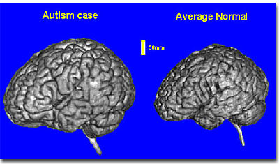
Autistic brains go their own way
Autism spectrum disorder (ASD) has been studied for many years, but there are still more questions than answers. For example, some research into the brain functions of individuals on the autism spectrum have found a lack of synchronization between different parts of the brain that normally work in tandem. But other studies have found the exact opposite - over-synchronization in the brains of those with ASD. A new study by Avital Hahamy and Prof. Rafi Malach of the Weizmann Institute’s Neurobiology Department, and Prof. Marlene Behrmann of Carnegie Mellon University, Pittsburgh, which was recently published in Nature Neuroscience, suggests that the various reports - of both over- and under-connectivity - may, in fact, reflect a deeper principle.
To investigate the issue of connectivity in ASD, the researchers analyzed data obtained from functional magnetic resonance imaging (fMRI) studies conducted while the participants were at rest. These had been collected from a large number of participants at multiple sites and handily assembled in the ABIDE database. “Resting-state brain studies are important,” says Hahamy, “because that is when patterns emerge spontaneously, allowing us to see how various brain areas naturally connect and synchronize their activity.” A number of previous studies in Malach’s group and others suggest that these spontaneous patterns may provide a window into individual behavioral traits, including those that stray from the norm.
In a careful comparison of the details of these intricate synchronization patterns, the researchers discovered an intriguing difference between the control and ASD groups: The control participants’ brains had substantially similar connectivity profiles across different individuals, whereas those with ASD showed a remarkably different phenomenon. These tended to display much more unique patterns - each in its own, individual way. The researchers realized that the synchronization patterns seen in the control group were “conformist” relative to those in the ASD group, which they termed “idiosyncratic.”
The researchers offer a possible explanation for differences between the synchronization patterns in the autism and control groups: They might be a product of the ways in which individuals in the two groups interact and communicate with their environment. Hahamy: “From a young age, the average, typical person’s brain networks get molded by intensive interaction with people and the mutual environmental factors. Such shared experiences could tend to make the synchronization patterns in the control group’s resting brains more similar to each other. It is possible that in ASD, as interactions with the environment are disrupted, each one develops a more uniquely individualistic brain organization pattern.”
Autistic Brains Show Too Many Connected Neurons
Autism may be a disorder of hyper-connectivity in the brain, according to a study that found children with the condition have too many synapses, the points where neurons connect and communicate with each other.
The study, published yesterday in the journal Neuron, suggests that a dysfunction in the brain doesn’t prune the neurons during development, as happens in most people. Researchers from Columbia University Medical Center examined tissue from the brains of children who had died, including those with and without autism.
Autism disorders are characterized by indifference to social engagement, communication difficulties and repetitive behaviors. There is no cure or single known cause, though studies have suggested a range of potential biological and environmental starting points. The finding adds a new pathway that may be targeted by a drug, researchers said.
“This is an important finding that could lead to a novel and much-needed therapeutic strategy for autism,” said Jeffrey Lieberman, chairman of psychiatry at Columbia University Medical Center, in a statement. He wasn’t involved in the study.
The researchers examined brain tissue from 26 children and adolescents ages 2 to 20 who had autism, and from 22 more who didn’t. The tissue was from a region of the brain involved in social and communication processes, and implicated in autism. They counted the number of spines extended from the neurons, each of which connect with another neuron by synapse.
 The researchers emphasize that this explanation is only tentative; much more research will be needed to fully uncover the range of factors that may lead to ASD-related idiosyncrasies. They also suggest that further research into how and when different individuals establish particular brain patterns could help in the future development of early diagnosis and treatment for autism disorders.
###
Prof. Rafael Malach’s research is supported by the Murray H. and Meyer Grodetsky Center for Research of Higher Brain Functions, which he heads; and the Friends of Dr. Lou Siminovitch. Prof. Malach is the recipient of the Helen and Martin Kimmel Award for Innovative Investigation; and he is the incumbent of the Barbara and Morris L. Levinson Professorial Chair in Brain Research.
A problem with the way the brain develops may leave autistic children with too many connections among their brain cells, or neurons. This might make them vulnerable to overstimulation and contribute to their autism symptoms, new research suggests.
During infancy, the number of synapses - the connections that allow neurons to send and receive information - grows rapidly. Later on, during childhood and adolescence, these connections are pruned back in response to learning and interacting with the environment.
In the study, published yesterday in the journal Neuron, researchers from Columbia University Medical Center and colleagues at other institutions looked at the temporal lobe of the brain, an area involved in communication and social behavior. The brains of older children with autism had a greater number of connections in the temporal lobe than the brains of their peers without autism.
Less Pruning in Brains of Kids with Autism
Researchers examined tissue taken from the brains of 26 deceased children between the ages of 2 and 19, half with autism and half without. The researchers counted the dendritic spines, which are the protrusions at the ends of neurons that receive signals across the synapses.
At younger ages, the two groups of children had a similar number of spines. Among typically-developing children, the number of synapses dropped by 41 percent as they grew older. In children with autism, the number decreased by only 16 percent.
Because the younger children had a similar number of connections in their brains, researchers suggest that the problem is not an overproduction of synapses, but instead trouble pruning back the excess.
“While people usually think of learning as requiring formation of new synapses, the removal of inappropriate synapses may be just as important,” the study’s senior investigator, David Sulzer, a professor of neurobiology at Columbia, said in a press statement.
The Weizmann Institute of Science in Rehovot, Israel, is one of the world’s top-ranking multidisciplinary research institutions. Noted for its wide-ranging exploration of the natural and exact sciences, the Institute is home to scientists, students, technicians and supporting staff. Institute research efforts include the search for new ways of fighting disease and hunger, examining leading questions in mathematics and computer science, probing the physics of matter and the universe, creating novel materials and developing new strategies for protecting the environment.
###
Yivsam Azgad
.(JavaScript must be enabled to view this email address)
###
Journal - Nature Neuroscience